Locate Us
- Doctor's Care Polyclinic & Diagnostic Centre, 168/299, Hakim Para, Opp. Ushasi Bhawan, Jalpaiguri-735101, West Bengal, India
- +91 97359 20690 / +91 97331 48734
- info@drrajatkrbhattacharjee.com
Laparoscopic (keyhole) surgery uses a laparoscope, a slim instrument with a camera and light at its tip, passed through small cuts in the abdomen. The surgeon operates while watching a high-definition monitor, so there is no need for a large open incision. For patients looking for the best laparoscopic surgery in North Bengal, this usually means less pain, smaller scars, a shorter hospital stay and a quicker return to work.
Dr. Rajat Kumar Bhattacharjee offers basic and advanced laparoscopic surgery in Jalpaiguri, along with diagnostic and therapeutic endoscopy, abdominal cancer surgery and laser proctology. Not sure whether keyhole surgery is right for you? Book a consultation at Doctor’s Care Polyclinic, Jalpaiguri.
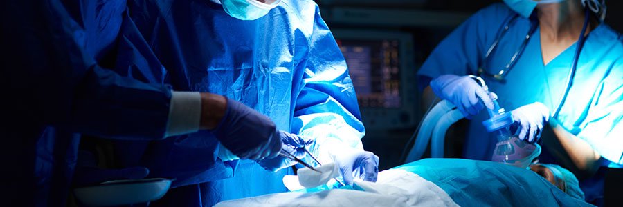
Compared to traditional open surgery, patients often experience less pain, a shorter recovery, and less scarring with laparoscopic surgery.
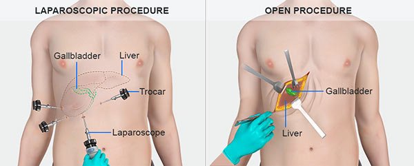
A cholecystectomy is a surgical procedure to remove your gallbladder — the small, hollow organ that stores bile for your digestive system. Gallbladder removal surgery is a common treatment for many types of gallbladder disease. This is because the downsides of having your gallbladder removed are generally fewer than those of the diseases it treats. You can live a healthy life without a gallbladder.
The operation is done the following way:
The gallbladder is then removed using the laparoscope and other instruments.
Common symptoms may include:
Symptoms usually last for short periods of time and often occurs after fatty meals which stimulate the gallbladder to contract.
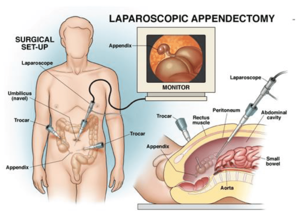
An appendectomy is a surgery to remove the appendix. It’s the main treatment for an inflamed appendix, commonly known as appendicitis.
The appendix is a small, tube-shaped pouch that’s attached to your intestines on the lower right side of your abdomen. Its exact purpose is unknown. Your body can still function without it.
It’s important to seek treatment right away if you’re having symptoms of appendicitis, such as:
If you don’t receive treatment for appendicitis, your appendix can rupture (burst).
A ruptured appendix can release bacteria and other toxins into your abdominal cavity. This can lead to a longer hospital stay and could be life threatening.
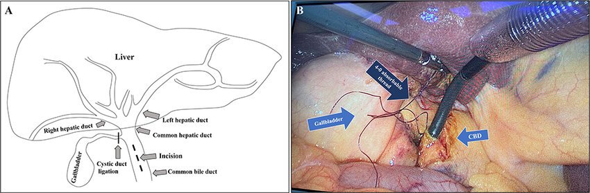
Laparoscopic cholecystectomy, also known as minimally invasive cholecystectomy, is performed through 4 small incisions with use of a camera to visualize the inside of the abdomen and long tools to remove the gallbladder. Surgery is done under anesthesia, and patients are asleep throughout the entire procedure.
Laparoscopic cholecystectomy is a safe and effective treatment for most children diagnosed with biliary disease. Although it takes longer to perform than open cholecystectomy does, it results in less postoperative narcotic use and a shorter hospital stay, as has been the case in the adult literature.
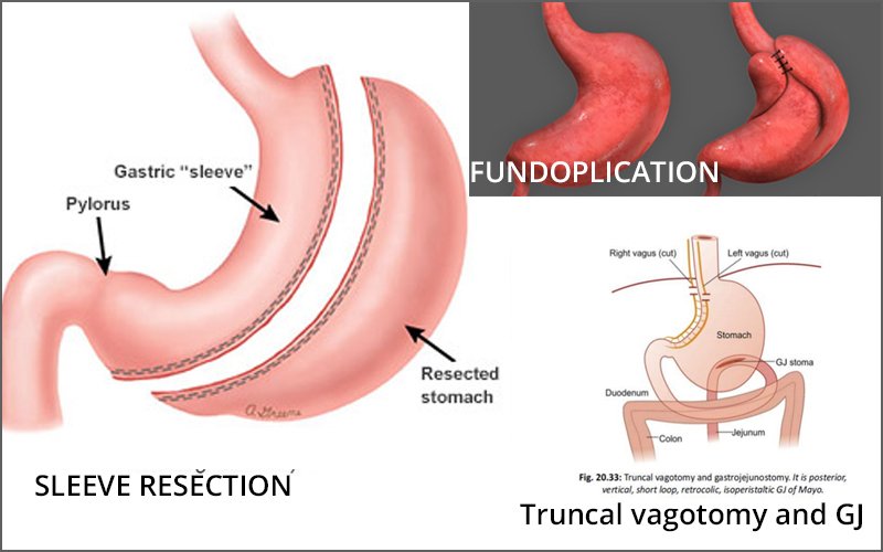
You might stay on IV fluids and nutrition at first, or you might begin with a clear liquid diet and gradually transition to a soft diet before going home. Occasionally, it can take several weeks for your stomach to start working again after surgery. In this case, you may continue to be fed through a tube or an IV.
Risks: Surgical procedures, including gastrojejunostomy, involve inherent risks like bleeding, blood clots, organ damage, and excessive internal scar tissue formation.
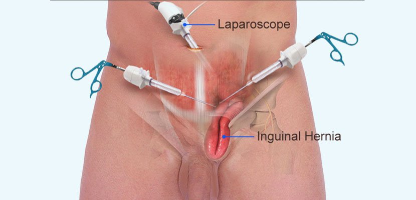
Laparoscopic hernia repair is a minimally invasive surgical technique used for repairing many types of hernias. This procedure uses three to four small incisions and a tool called a laparoscope, or a thin tube with a camera on the end, to guide the surgeon. In some cases, mesh may be used to hold the protruding tissues in place. A laparoscopic hernia repair typically takes one hour or less to complete. Dr. Rajat Kumar Bhattacharjee performs Hernia Surgery in Jalpaiguri.
Pre-procedure instructions will be explained to patients by the surgeon. It may be necessary to stop eating and drinking for at least eight hours before the procedure. Patients taking prescription drugs should ask their doctor if they should continue taking their medications as usual on the days leading up to surgery. This is because certain medications can affect the body’s ability to clot blood, which can lead to complications during and/or after surgery.
Minimally invasive, minimal incision techniques provide the following benefits to patients:
Lower risk of infection
Smaller incision size
Shorter operating room (OR) times
Earlier return to work and daily activities
Less pain and shorter recovery time for some patients
Additionally, if necessary, laparoscopic techniques allow surgeons to repair multiple hernias using the same incisions.
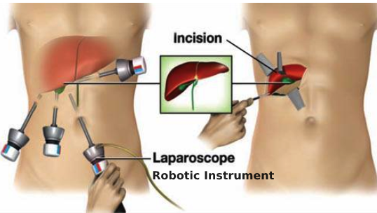
A. The Condition. You may have a cyst in your liver. These can be the result of an infection. The cyst is removed or destroyed with a laparoscopic hepatic cystectomy.
B. Symptoms. You may have the following symptoms:
C. Laparoscopic Hepatic Cystectomy. The surgeon will make about 3-4 small incisions in your abdomen. A port (nozzle) is inserted into one of the slits, and carbon dioxide gas inflates the abdomen. This process allows the surgeon to see inside of your abdomen more easily. A laparoscope is inserted through another port. The laparoscope looks like a telescope with a light and camera on the end so the surgeon can see inside the abdomen. Surgical instruments are placed in the other small openings and used to remove or destroy the cyst within your liver. Your surgeon will use a special instrument which cauterizes the liver as it is cut, so that there will be minimal bleeding. If a liver specimen is removed, then it is extracted from your abdomen in a plastic bag by enlarging one of the small incisions. After this has been accomplished, the carbon dioxide is released out of the abdomen through the slits, and then these sites are closed with sutures or staples, or covered with glue-like bandage and steri-strips. When you wake up, you may have a temporary drain or rubber tube exiting out of your skin.
D. Before Your Operation. Laparoscopic hepatic cystectomy usually is an elective procedure. The preoperative evaluation might include blood work, urinalysis, an abdominal ultrasound, and an abdominal CT scan. In some cases, endoscopy and/or x-rays of your bile duct may be performed. If you smoke, then you should stop immediately. If you are taking blood thinners (for example, aspirin, coumadin, or Plavix), then you will need to stop these one week prior to your procedure. Your surgeon and anesthesia provider will review your health history, medications (including blood thinners), and options for pain control.
E. Your Recovery. You usually can go home in 1 – 2 days after a laparoscopic hepatic cystectomy. You may need to wait until your bowels start working. You will be given medication for pain, and you may need to take antibiotic pills. If you have a drain, then your doctor and/or nurse will instruct you in the care for the drain. You should limit your activity to light lifting (no more than 15 lb) for one month.
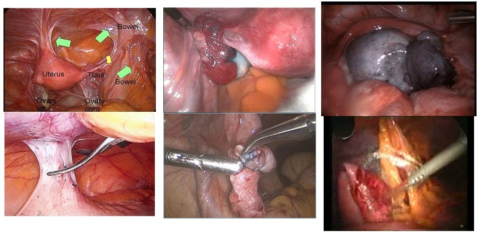
A gynecologic laparoscopy is a procedure for some tests and treatments. Small cuts (incisions) are made in the belly (abdomen). Then, a slime telescope (laparoscope) is used to see inside the body. Some conditions that may be found (detected) or treated are:
Any tissue from the biopsy or a removed cyst will be tested. You will be contacted with the results. This may take up to 2 weeks.
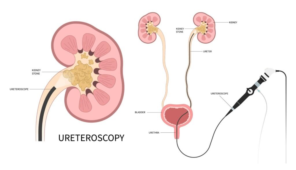
Kidney stones are one of the most common reasons people in Jalpaiguri and across North Bengal see a surgeon for sudden pain in the back or side. A stone forms when minerals and salts in the urine become too concentrated and crystallise into a hard lump. Some stones stay in the kidney without causing trouble; others move down the ureter and block the flow of urine.
Many small stones pass naturally with plenty of fluids and the right medicines. A stone needs to be removed when it is too large to pass, keeps coming back, causes infection or starts to damage the kidney. Today most stones can be treated with minimally invasive techniques, so a large open cut and a long hospital stay are rarely needed.
This section explains the main types of kidney stones, the symptoms to watch for, and when laparoscopic (keyhole) surgery is the right choice.
A stone sitting quietly in the kidney may cause no symptoms at all. Trouble usually starts when it moves into the ureter, the narrow tube that carries urine from the kidney to the bladder. Common signs are:
Stones form when urine carries more minerals than it can dissolve, so crystals begin to build up. There is rarely a single cause; usually several factors combine:
Most stones are treated first with shock-wave lithotripsy or with endoscopic and laser procedures that reach the stone through the urinary tract (ureteroscopy) or through a small opening in the back (PCNL). Laparoscopic surgery is chosen when these are unlikely to work, for example:
The operation is done under general anaesthesia. The surgeon makes three or four small cuts of about 5–10 mm in the abdomen, uses a camera to find the stone, removes it and stitches the opening in the kidney or ureter. A thin, soft tube (a DJ stent) is usually left inside to help urine drain while the area heals; it is taken out in a short follow-up procedure a few weeks later. Most patients go home within two to three days.
Compared with open surgery, the keyhole approach usually means:
If you have repeated episodes of kidney pain, or a scan has shown a large stone, book a consultation with Dr. Rajat Kumar Bhattacharjee in Jalpaiguri. After reviewing your reports, the doctor will explain whether medicine, an endoscopic procedure or laparoscopic surgery is the best option for you.

Basic & Advanced Laparoscopic Surgery, Specialization in Diagnostic & Therapeutic Endoscopy, Abdominal Cancer Surgery, Laser Proctology.